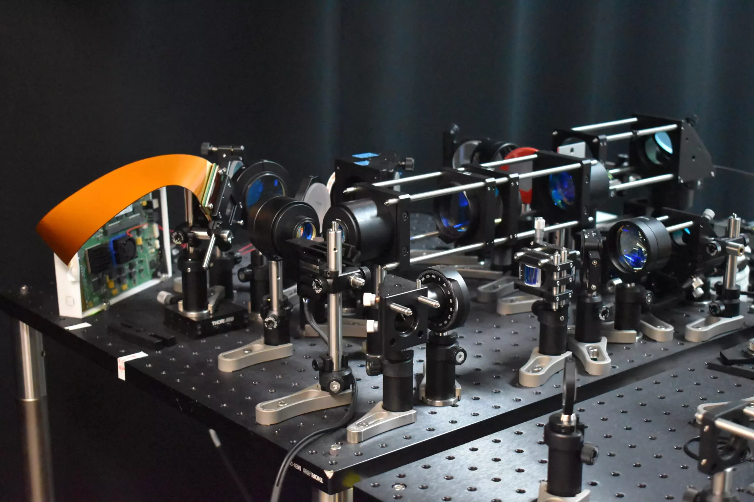
The latest advancements in two-photon fluorescence microscopy have led to the development of a new microscope that is capable of capturing high-speed images of neural activity at cellular resolution. This new approach allows researchers to image neurons much faster and with less harm to brain tissue than traditional two-photon microscopy, opening up new possibilities for studying the dynamics of neural networks in real time.
The new two-photon fluorescence microscope incorporates a novel adaptive sampling scheme and replaces traditional point illumination with line illumination. This innovative approach enables researchers to conduct in vivo imaging of neuronal activity in a mouse cortex at speeds ten times faster than traditional methods, while also reducing the laser power on the brain more than tenfold. The use of line illumination allows for a larger area to be excited and imaged at once, significantly speeding up the imaging process.
By providing a tool that can observe neuronal activity in real time, this new technology has the potential to significantly impact neuroscience research. Researchers could use this microscope to study the dynamics of neural networks during fundamental brain functions such as learning, memory, and decision-making. Furthermore, the ability to monitor neural activity in real time could help researchers better understand and treat neurological diseases such as Alzheimer’s, Parkinson’s, and epilepsy.
One of the key features of the new microscope is its adaptive sampling scheme, which uses a digital micromirror device (DMD) to dynamically shape and steer the light beam. This allows for precise targeting of active neurons, reducing the total light energy deposited to the brain tissue and lowering the risk of potential damage. By only imaging neurons of interest, the new technology minimizes harm to living tissue while providing detailed and high-speed imaging capabilities.
The researchers demonstrated the capabilities of the new microscope by imaging calcium signals in living mouse brain tissue at a speed of 198 Hz, showcasing its ability to monitor rapid neuronal events. In the future, the researchers plan to integrate voltage imaging capabilities into the microscope to provide a direct and rapid readout of neural activity. They also aim to enhance the user-friendliness and reduce the size of the microscope to make it more accessible for neuroscience research.
Overall, the advancements in two-photon fluorescence microscopy represent a significant leap forward in the study of neural activity. The new microscope technology offers the potential to gain deeper insights into how neurons communicate in real time, leading to a better understanding of brain function and neurological diseases. This innovative approach paves the way for exciting new discoveries in neuroscience research and opens up new avenues for studying the complexities of the brain.


Leave a Reply