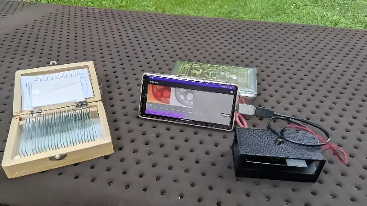
In the ever-evolving realm of microscopy, innovation continues to reshape how we explore and understand the microscopic world. A striking breakthrough has emerged from researchers at the Tokyo University of Agriculture and Technology: a smartphone-based digital holographic microscope. This compact and cost-effective technology promises to democratize access to precise 3D measurements across diverse applications, enhancing educational experiences and expanding diagnostic capabilities, particularly in underserved regions.
Before delving into the specifics of this groundbreaking microscope, it’s essential to grasp the fundamental principles of digital holographic microscopy (DHM). Unlike conventional microscopy, DHM utilizes the interference of light waves to generate detailed holograms, which are then digitally reconstructed to provide three-dimensional insights into various samples. This method allows researchers to visualize intricate internal structures and surface details without altering the specimen—a crucial advantage for biological samples.
However, traditional DHMs often come with significant drawbacks. They typically incorporate complex optical setups coupled with powerful personal computers for data processing, which severely limits their portability and usability in varied environments. Many current systems are impractical for fieldwork or in any setting without reliable access to advanced computing resources.
Yuki Nagahama, the leading researcher of this pioneering project, highlights a transformative shift in design philosophy. By utilizing a lightweight optical system fabricated through 3D printing alongside the computational power of smartphones, this new microscope has successfully minimized both size and cost. “This simplifies the technology,” Nagahama explains, citing the potential of this portable device for educational purposes in schools and for medical diagnostics in developing countries, where access to advanced equipment is limited.
This smartphone integration marks a crucial turning point. Previous smartphone-based DHMs struggled with reconstruction in real time, primarily due to the constraints of smartphone processing abilities. However, through innovative techniques like band-limited double-step Fresnel diffraction, researchers have navigated these limitations. This approach optimizes data processing, achieving faster reconstruction speeds, making real-time analysis not only possible but practical.
Applications and Potential Impact
The implications of this technology are significant. By enabling real-time hologram reconstruction directly on smartphones, a broad spectrum of applications becomes feasible. For instance, in medical diagnostics, this microscope could facilitate on-site evaluations for conditions like sickle cell disease, providing rapid responses that could save lives in settings where immediate medical attention is crucial.
Moreover, its portability opens doors for educational uses, allowing students to engage with live specimens both in the classroom and at home. The ability to zoom in on holographic images using simple swipe gestures adds an interactive dimension to learning, transforming static education into a dynamic exploration of life sciences.
Validation Through Testing
Researchers have rigorously evaluated their smartphone-based microscope by using test objects with known patterns. The results verified the system’s capability to accurately capture and reconstruct these patterns, showcasing its viability. Additionally, the microscope was successfully employed to image various biological samples, such as the cross-section of a pine needle, further illustrating its versatility.
These tests have confirmed that, thanks to the band-limited double-step Fresnel diffraction technique, the microscope can reconstruct holograms at astonishing speeds of up to 1.92 frames per second. While this may seem modest compared to traditional systems, it represents a monumental leap forward for portable devices, allowing nearly instantaneous observations of stationary subjects.
Looking ahead, the research team plans to leverage advancements in deep learning to refine the image quality produced by their system. One of the challenges faced by digital holographic microscopes is the occurrence of unintended artifacts during the hologram reconstruction process. By integrating deep learning algorithms, researchers aim to eliminate these artifacts, providing clearer and more reliable images, further enhancing the microscope’s usability.
The intersection of deep learning with optical microscopy could usher in a new era of precision imaging, making microscopy accessible not just in advanced research facilities but also in everyday educational and healthcare settings around the globe.
The development of a smartphone-based digital holographic microscope stands as a testament to the innovative potential rooted in merging traditional scientific methods with modern technology. By prioritizing accessibility, portability, and real-time functionality, researchers are not only expanding the horizons of microscopic analysis but are also making significant strides toward equitable healthcare and education. As the journey of this cutting-edge technology continues, it is keenly poised to transform how we visualize and analyze the unseen world surrounding us.


Leave a Reply