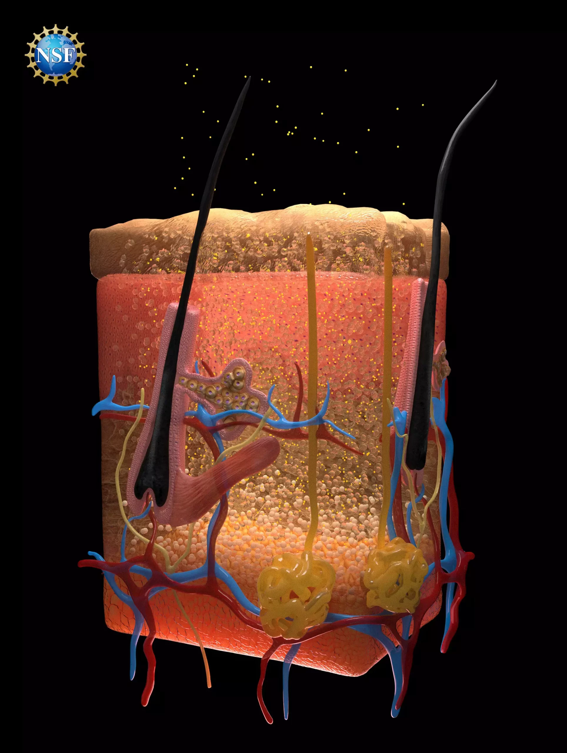
Recent advancements in medical diagnostics have unveiled a novel technique that could fundamentally alter how we visualize internal organs. Researchers from Stanford University have harnessed the power of optical transparency through innovative applications of food-safe dyes to render biological tissues transparent to visible light. This remarkable study, titled “Achieving optical transparency in live animals with absorbing molecules,” was published in the prestigious journal Science. The implications of this research could extend far beyond mere visualization; it may open new horizons in diagnosing injuries, monitoring digestive conditions, and detecting cancers.
The methodology employed in this study involves altering the optical properties of biological tissues through a topical application of dyes. At its core, the challenge lay in overcoming the scattering of light—a key reason why the human body appears opaque. Biological tissues consist of various materials with different refractive indices, causing light to scatter as it passes through. By matching these indices, the researchers could facilitate a smoother passage of light, thereby rendering the tissues transparent.
The pivotal substance in this research was identified as tartrazine, a commonly used food dye. This dye effectively raises the refractive index of biological fluids, aligning it with the refractive indices of surrounding tissues. This fascinating interplay between chemistry and optics highlights the researchers’ deep understanding of how light interacts with matter, allowing them to overcome long-standing limitations in biological imaging.
To validate their approach, the Stanford team conducted experiments on animal models, beginning with thin slices of chicken breast. By increasing the concentration of tartrazine, they observed a transition from opacity to clarity, as the refractive indices aligned perfectly. Following this success, the researchers applied the solution to living mice, rendering their skin transparent. This procedure showcased the blood vessels beneath the surface and demonstrated significant movement of internal organs such as the intestines, heart, and lungs.
These breakthroughs underscore not only the potential for enhanced medical imaging but also the possibility of real-time monitoring of biological functions. The ability to visualize processes at the micron level introduces unprecedented opportunities for medical diagnostics and research, allowing for better assessment of conditions that previously went unnoticed.
The real-world applications of this technology are vast and varied. From facilitating blood draws to improving laser treatments such as tattoo removal, the potential benefits touch multiple aspects of healthcare. In oncology, for instance, this technique could enhance the effectiveness of laser therapies by improving light penetration in deeper tissues, thereby increasing the accuracy of treatments for both cancerous and precancerous conditions.
Moreover, the reversibility of this technique is noteworthy. Upon rinsing the dye away, tissues swiftly recover their original opacity, ensuring that biological integrity remains intact. This characteristic is crucial for both clinical settings and experimental research, as it allows for repeated examinations without adverse effects on the subjects.
The journey from concept to application was fueled by a combination of theoretical insights and rigorous experimentation. The researchers leveraged foundational principles from optics, particularly focusing on concepts outlined in older textbooks, such as Kramers-Kronig relations and Lorentz oscillation. These equations provided a framework for predicting how varying dyes could impact the optical attributes of biological materials, ultimately guiding the selection of viable candidates for further testing.
Graduate researcher Nick Rommelfanger and postdoctoral researcher Zihao Ou played pivotal roles in transitioning these theories into practice, meticulously assessing various dyes for their optical compatibility. Their efforts underscore the importance of interdisciplinary collaboration in scientific discovery, bridging the gap between theory and innovation.
The findings from this study hold the promise of spawning a new field of study dedicated to matching dyes with biological tissues based on their optical properties. This pioneering approach not only expands our understanding of optics in a medical context but also opens the door for future research aimed at refining and improving this technology.
As Richard Nash, an NSF Program Officer, noted, the integration of traditional tools like ellipsometers in innovative applications can lead to profound discoveries in science. This research exemplifies how an unexpected marriage of basic principles and modern experimentation can yield groundbreaking results.
The advent of this optical transparency technique could herald a new era of medical imaging, empowering healthcare professionals with enhanced diagnostic tools. As the research community continues to explore and refine these methodologies, the potential benefits to patient care and medical understanding are not just promising—they are groundbreaking.


Leave a Reply