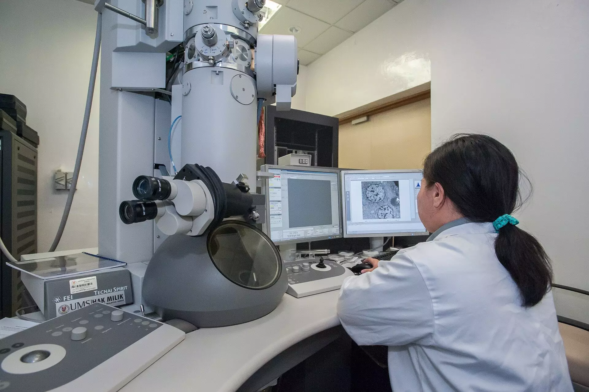
In an era marked by rapid technological advancements, a collaborative team from Trinity College Dublin has made a groundbreaking stride in imaging technology, promising to reshape the field of microscopy. By implementing a sophisticated imaging method that leverages high-end microscopes, this innovative approach significantly minimizes both the time and radiation exposure traditionally associated with electron microscopy. As the frontiers of science continue to expand, such advancements not only enhance research capabilities but also ensure the integrity of delicate samples, particularly in the realms of medicine and materials science.
The Challenge of Traditional Imaging Techniques
Current scanning transmission electron microscopes (STEMs) function by directing a concentrated electron beam at samples, capturing images pixel by pixel. This conventional method necessitates a predetermined amount of “dwell time” for each pixel, during which the microscope pauses to gather signals from the sample. While this process, reminiscent of classic film cameras, has served researchers well, it comes with significant drawbacks. The incessant bombardment of electrons can lead to damage, especially in sensitive biological tissues. As scientists explore increasingly delicate materials, the potential for sample destruction becomes a pressing concern.
It is crucial to recognize that the consistent exposure times across different areas of an image do not consider the variable nature of the sample features. The fixed dwell time might yield substantial data in some regions, while being superfluous and harmful in others. The result is an inefficient imaging process where valuable samples could degrade under harsh conditions, leading to compromised results.
Innovative Solutions: The Event-Based Detection System
The new method introduces a paradigm shift by moving away from the traditional fixed dwell-time model. Instead, the team has devised an event-based detection system, measuring the time taken to capture a set number of electron events. This revolutionary approach hinges on a mathematical theory that underscores the efficiency of the first electron detected at any given probing position. Intriguingly, while each additional electron provides diminishing returns in information, they all carry the same risk of damaging the sample.
This breakthrough capability allows scientists to “shut off” the electron illumination at the pinnacle of imaging efficiency, significantly reducing the number of electrons needed to produce high-quality images. By doing so, researchers can enjoy enhanced imaging without the hefty radiation cost. The mathematical insights supporting this strategy are as important as the practical implementation itself, paving the way for a new standard in microscopy.
From Theory to Practice: Tempo STEM Technology
While a theoretical advancement is impressive, its practical realization is what truly matters in scientific progress. To bring this innovative approach to fruition, the research team has patented a groundbreaking technology known as Tempo STEM, in collaboration with IDES Ltd. This device integrates a sophisticated “beam blanker” that seamlessly shutters the electron beam once the desired imaging quality has been achieved. According to Dr. Lewys Jones, a key figure in the research effort, this innovative capability represents an unprecedented leap in microscope functionality.
This advancement not only streamlines the imaging process but directly addresses the critical issue of radiation exposure, enabling microscopists to operate within a framework where excessive exposure becomes obsolete. Dr. Jon Peters, the lead author on the paper, highlights the misconception regarding the harmlessness of electrons. When accelerated to 75% the speed of light and directed at fragile biological specimens, even electrons can wreak havoc, yielding unusable or misleading images.
Broader Implications for Science and Research
The implications of this innovation extend far beyond the boundaries of microscopy. Researchers in various fields including nanotechnology, biology, and materials science stand to benefit immensely from improved imaging techniques. This advancement not only enhances the quality of research output but also stimulates further explorations into the nano-world, where precision is paramount and sample integrity is non-negotiable.
In a landscape where scientific endeavors increasingly rely on precision imaging, the introduction of Tempo STEM and its associated techniques represents a vital step forward. The integration of imaginative mathematical models with cutting-edge technology stands as a testament to the power of interdisciplinary collaboration in revolutionizing scientific tools. With imaging methods that promise not just higher quality, but also safety for delicate samples, the future for researchers appears incredibly bright.


Leave a Reply