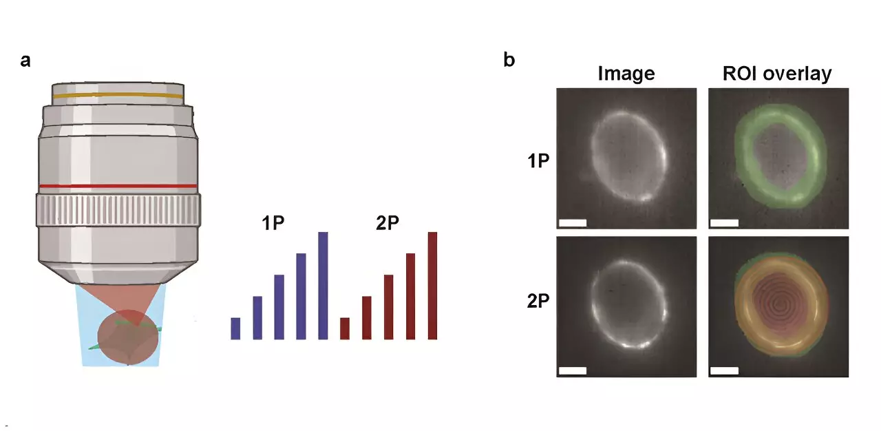
In the realm of neuroscience, the investigation of neural circuitry is crucial for deciphering how the brain processes information. Genetically encoded voltage indicators (GEVIs) have emerged as powerful tools in this quest, enabling researchers to visualize electrical activity within neurons. These indicators have revolutionized our understanding of neuronal communication, yet the debate concerning the effectiveness of various imaging techniques continues. A recent study conducted by Harvard University researchers provides an essential comparison of one-photon (1P) and two-photon (2P) voltage imaging, revealing key insights regarding their advantages and disadvantages.
The study published in Neurophotonics meticulously examined the optical and biophysical limits inherent in both 1P and 2P imaging methods. The researchers assessed the brightness and voltage sensitivity of widely used GEVIs under distinct illumination conditions, providing empirical data critical for practical applications. An essential aspect of their investigation was the fluorescence behavior with varying depths in the mouse brain, an element of vital importance for in vivo studies.
To quantify the implications of their findings, the research team established a model aimed at predicting the measurable cell populations based on various parameters. These included the properties of the imaging reporter, the specific imaging configurations employed, and the targeted signal-to-noise ratio (SNR). Such quantification is crucial for setting benchmarks for future experiments.
One striking conclusion of the study highlights the substantial energy requirements associated with 2P voltage imaging. The researchers found that 2P excitation necessitates an illumination power around 10,000 times greater per cell than its 1P counterpart to generate comparable photon count rates. This discrepancy results in practical concerns, particularly regarding tissue photodamage and shot noise, complicating the in vivo application of 2P imaging.
For instance, when measuring neuronal activity in the mouse cortex using the JEDI-2P indicator, the team determined that with a target SNR of 10, limitations in laser power (set at 200 mW) restricted the ability to record from more than approximately 12 neurons located deeper than 300 micrometers. Such restrictions highlight the scarcity of viable strategies for robust in vivo imaging that can capture numerous neurons simultaneously at deeper brain layers.
The researchers concluded that current GEVIs exhibit modest voltage sensitivity and impose stringent photon-count needs, thereby presenting significant challenges for effectively employing 2P voltage imaging in live subjects. Future advancements must either enhance 2P GEVIs or pioneer entirely new imaging modalities to facilitate high SNR readings for large populations of neurons, especially at considerable depths in the brain.
This study emphasizes the intricate balance between 1P and 2P voltage imaging techniques and underlines the pressing need for innovation within imaging technologies. The journey toward overcoming these obstacles holds promise for significantly enhancing our comprehension of neural circuits, driving further scientific inquiry and discovery in the field of neuroscience. The findings from this research will undoubtedly serve as a guiding framework for subsequent advancements in voltage imaging, pushing the boundaries of our understanding of the brain’s complexities.


Leave a Reply